Researchers have recently stumbled upon an astonishing revelation about the organization of muscle fibers in Parophidion vassali, a fish species inhabiting the Mediterranean Sea. This groundbreaking discovery has the potential to reshape our understanding of muscle contraction dynamics.
Eric Parmentier and Marc Thiry, scientists from the University of Liège, have unveiled an extraordinary arrangement of muscle fibers in Parophidion vassali, which could revolutionize our comprehension of muscle contraction. This distinctive network-like configuration of myofibrils within the muscle fiber allows for swift contractions while preserving strength. Further investigation is required to fully grasp the implications and functionality of this novel muscle fiber structure.
The history of skeletal muscle description traces back to Antoni van Leeuwenhoek, a Dutch biologist and pioneer in cell biology and microbiology. In 1712, van Leeuwenhoek utilized a portable single-lens microscope to provide the first account of muscle fibers found in a whale, as documented in an article published in the Philosophical Transactions of the Royal Society. In 1840, anatomist William Bowman offered a more precise depiction of muscles, noting the delicate and intricate “alternately light and dark lines” he observed. Subsequent studies have enhanced our understanding of muscle fibers, including the identification of their molecular components and the proposal of the muscle contraction model by biophysicist Andrew Huxley in 1957. Over the past three centuries, numerous studies across different animal groups have consistently revealed the preserved general organization of striated muscle fibers.
Nonetheless, the proportion of cellular components within each fiber can vary, imparting distinct contraction properties. For instance, muscles responsible for forceful contractions contain myofibrils with a less developed sarcoplasmic reticulum. Conversely, muscles capable of rapid contractions have fewer myofibrils but an abundance of sarcoplasmic reticulum and mitochondria. The sonic muscles of fish exhibit the highest contraction speeds, producing sounds through muscles that contract at a frequency of 100 to 300 Hz, equivalent to 100 to 300 contraction/relaxation cycles per second.
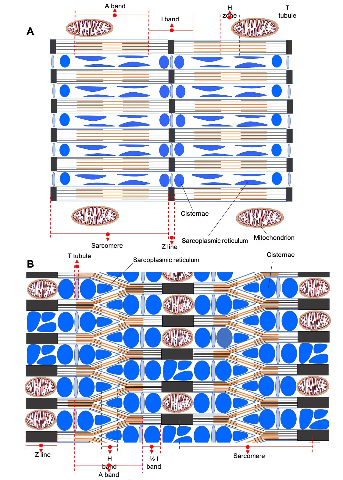
In a recent collaborative study conducted by the Functional and Evolutionary Morphology Laboratory and the Cell and Tissue Biology Laboratory, a new myofibril arrangement within the fibers of Parophidion vassali’s sonic muscle was unveiled. Instead of a parallel arrangement, the myofibrils form an extensive network within the muscle fiber. According to Professor Eric Parmentier, director of the Functional and Evolutionary Morphology Laboratory, this unique muscle fiber design enables rapid contractions while maintaining strength. The scarcity of myofibrils and the significant volume occupied by the sarcoplasmic reticulum favor swift contraction in these fibers.
Professor Marc Thiry, director of the Cell and Tissue Biology Laboratory, explains that the network structure of myofibrils allows for increased cross-bridging between myosin heads and actin myofilaments, enhancing strength in fast muscles. Additionally, the unusually arranged mitochondria within the Z striae provide the necessary energy for producing long-lasting sounds, with these fibers exhibiting a longer Z striae length (700 nm compared to the conventional range of 70 to 150 nm).
This unique organization of striated muscle fibers, previously unreported in scientific literature, holds the potential to combine muscular strength and speed. Further studies are required to fully comprehend its mechanisms and determine any molecular adaptations within these muscles.
Classic skeletal muscle: Longitudinal section. Credit: Marc Thiry/University of Liège
About Muscle Fibers:
Striated skeletal muscle fibers, constituting the fundamental units of voluntary muscles, exhibit parallel bundles of contractile elements known as actin and myosin myofilaments. These fibers appear as alternating light and dark bands, forming transverse striations in the longitudinal section under an optical microscope. The Z striation, dividing the light band, marks the midpoint. A sarcomere, the contractile unit of a myofibril, represents the portion between two Z striae. Many sarcomeres are arranged sequentially, forming a myofibril.
Occupying a significant cell volume, myofibrils are surrounded by cisternae of the sarcoplasmic reticulum, which stores calcium crucial for muscle contraction. Mitochondria, the primary source of ATP, the energy required for muscle contraction, are located in proximity to the myofibrils.
Table of Contents
Frequently Asked Questions (FAQs) about muscle fiber structure
What is the significance of the newly discovered muscle fiber structure in Parophidion vassali?
The newly discovered muscle fiber structure in Parophidion vassali is significant because it challenges our current understanding of muscle contraction. This unique network-like configuration of myofibrils within the muscle fiber allows for rapid contractions while retaining strength. It could potentially revolutionize our knowledge of muscle function.
How was the discovery of the muscle fiber structure made?
The discovery of the muscle fiber structure was made by researchers at the University of Liège, Eric Parmentier and Marc Thiry. They conducted a study on the Mediterranean fish species, Parophidion vassali, and observed the unique arrangement of myofibrils within the muscle fibers using advanced imaging techniques.
How does this muscle fiber structure differ from traditional muscle fiber organization?
Unlike traditional muscle fiber organization seen in most vertebrate animals, where myofibrils are arranged in parallel, the muscle fibers in Parophidion vassali exhibit a lattice-like network arrangement. This novel structure allows for rapid contractions while maintaining strength, which is different from the conventional organization observed in other species.
What are the potential implications of this discovery?
The discovery of this unique muscle fiber structure has the potential to revolutionize our understanding of muscle contraction and could have implications in various fields such as biomechanics, physiology, and evolutionary biology. Further research is needed to fully comprehend the functional implications and potential adaptations associated with this muscle fiber structure.
Are there any other species with similar muscle fiber structures?
To date, this type of muscle fiber structure has not been described in scientific literature, making it a unique finding. However, further studies may reveal if there are any similar muscle fiber structures in other species or if Parophidion vassali possesses any specific adaptations related to its muscle function.
More about muscle fiber structure
- University of Liège
- Functional and Evolutionary Morphology Laboratory
- Cell and Tissue Biology Laboratory


