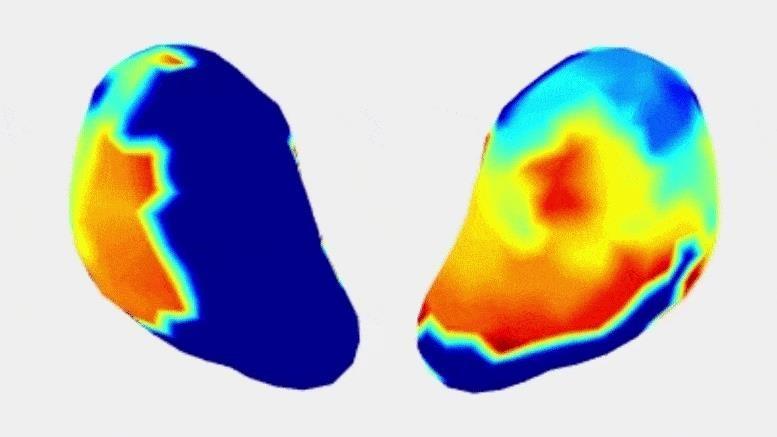
New Technology Maps Uterine Contractions With Incredible Accuracy
Scientists from Washington University School of Medicine have invented a special imaging device that can map out the strength and position of uterine contractions during childbirth. This means it gives us more accurate reading than other tools out there, which simply tell us if there is a contraction happening or not. The technology is based on the same type of images we use to look at our hearts. Best part? It can do all this without any invasiveness!
Today, March 14, a study was published in the journal Nature Communications. This study included 10 people who were having a baby.
Researchers have recently developed a special imaging technology that can capture and measure 4-dimensional maps of uterine contractions. This upgrade in measuring labor contractions could possibly help experts care for pregnant women with higher risks and even find ways to prevent premature birth, which is something that affects about 10% of pregnancies around the world.
Researchers at Washington University School of Medicine have just created a cool 3D map technology that shows how your uterus contracts in real-time! This new tech could help understand how healthy labor works, and detect if there are any issues like preterm labor or the labor stopping. In the video clip, you can see both sides of one contraction during normal labor.
When a baby is born, the uterus contracts in order to push out the baby. To measure these contractions, scientists use something called electromyometrial imaging (EMMI). This technology helps us know what types of early contractions lead to a preterm birth, which means the baby arrives earlier than expected. If contractions are not normal they can stop labour completely and then a C-section might have to happen. Preterm births and C-sections can cause issues for both parents and babies like birth injuries or even death. These injuries can even cause long-term health problems later on in life for the child.
A research team created a technology that tracks heart contractions, but they also found out it can also be used to measure uterine contractions. The thing is, uterine contractions are not as predictable or consistent as the heart contractions. The starting point and direction of those labor contractions may change from time to time even for the same person. What’s more, the team noticed that these baby-producing muscle squeezes don’t start in any particular area so this imaging tech can track changes through every contraction.
The researchers looked at different types of moms who were having babies. Some had never given birth before and some had already done so in the past. Moms who hadn’t had babies yet had contractions that lasted longer with more changes during their labor, suggesting that their bodies might remember an old labor experience from a previous one. But for mothers who had already experienced childbirth before, their uteruses understood better how to produce efficient and effective contractions.
The Professor, Wang, suggests some medical uses of EMMI. These include knowing when a contraction is helpful or not for predicting when a baby will be born early, watching labor contractions in real-time to get medicines and treatments right so the delivery doesn’t have any issues, and keeping an eye on after-birth contractions to avoid bleeding.
Scientists are looking for new ways to treat problems related to the uterus, like painful periods and endometriosis, without using medicines. This could be done through electricity or other treatments that can help with contractions in the uterus.
Dr. Wang wants to find out if the contractions of a woman in labor are strong enough to lead her to giving birth. His team was given money by the National Institutes of Health (NIH) which they will use to make a chart showing what normal labor contractions look like.
This program is aiming to capture how everything looks like during a normal birth, whether it’s the first time around or multiple times. This is all new information so we need collect facts and form something called an “atlas” that we can refer to as our reference point.
In places with limited resources, new imaging technology can make labor and childbirth safer. To make it more accessible to people, they are going to use cheaper and portable ultrasound imaging instead of the expensive MRI scans that aren’t available in many parts of the world. A team is working on this project with funding support from the Bill & Melinda Gates Foundation. They are coming up with single-use electrodes and wireless transmitters with help from Chuan Wang and Shantanu Chakrabartty who both work at Washington University.
Yong Wang said they are making a super low-price machine to be used in places with less money. This machine uses printed and disposable electrodes together with a wireless transmitter that would keep the cost down.
A group of scientists who are from different universities in the United States published a research paper called “Noninvasive electromyometrial imaging of human uterine maturation during term labor” on 14th March 2023. The research is about looking at how a baby grows before it is born. This study was published in Nature Communications and has an access code of 10.1038/s41467-023-36440-0 which can be used to find more information.
This work was funded by different organizations like March of Dimes, National Institute of Child Health and Human Development of the NIH, Burroughs Wellcome Fund Preterm Birth Initiative, and Bill & Melinda Gates Foundation. This funding was used to analyze the application of atosiban in a test-tube baby program as part of a fresh embryo transfer cycle.


