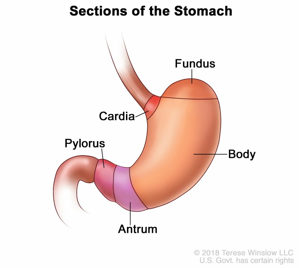
The stomach is a muscular, sac-like organ in the gastrointestinal tract of humans and many other animals, including several invertebrates. The stomach has a dilated structure and functions as a temporary storage reservoir for food. It is anatomically located between the esophagus and the small intestine. The main anatomical features of the stomach include the fundus, body, and pylorus.
The human stomach typically holds about one liter of material at any given time. It receives food from the esophagus via peristalsis and releases it into the duodenum through peristaltic contractions and controlled relaxation of an internal valve called the pyloric sphincter. These movements are coordinated by a complex interplay of local hormones like gastrin, motilin, cholecystokinin (CCK), ghrelin, secretin, vasoactive intestinal peptide (VIP), histamine, somatostatin; as well as nervous system inputs from both intrinsic nerves like gastric parasympathetic nerve fibers (vagus nerve) and extrinsic nerves from sympathetic ganglia like celiac ganglion or solar plexus. Various types of specialized cells line all surfaces of the stomach: mucous cells secrete digestive enzymes and mucus that protect epithelial surfaces; endocrine cells secrete various hormones that help coordinate digestive processes; enterochromaffin-like (ECL) cells secrete histamine in response to gastrin stimulation which increases acid secretion; chief cells secrete digestive enzymes; parietal cells secrete hydrochloric acid (HCl); G cellsecrete gastrin in response to peptides in chyme; D cellssynthesize and release somatostatin in response to high concentrations of amino acids which inhibits gastrin secretion among other things. All these various cell types together with contraction/relaxation patterns mediated by smooth muscle constitute what is known as the migrating motor complex responsible for gut motility patterns that sweep unabsorbed materials towards evacuation.
Mucous membranes line all surfaces inside the stomach except for areas exposed to luminal contents where there are only epithelial layers backed up by a layer connective tissue rich in blood vessels called lamina propria underneath it all. In most mammals including humans there are three distinct regions on these mucosal surfaces:
* Cardia: closest to where the esophagus opens into stomach
* Fundus: dome shaped area above cardia
* Body: main central region below fundus
These regions can be further subdivided into four quadrants based on two perpendicular axes running through center of each: right upper quadrant(RUQ), left upper quadrant(LUQ), right lower quadrant(RLQ), left lower quadrant(LLQ). Each surface inside these regions have different names too e.g., greater curvature vs lesser curvature referring to concave vs convex shape formed by topography of organs when viewed along longitudinal axis respectively; or anterior wall vs posterior wall referring to front vs back side when viewed along transverse/coronal axis respectively etcetera. For example “posterior wall” would refer to back side if you were looking at someone standing upright whereas “dorsal wall” would refer to same thing if you were looking at them lying down on their back etcetera.


